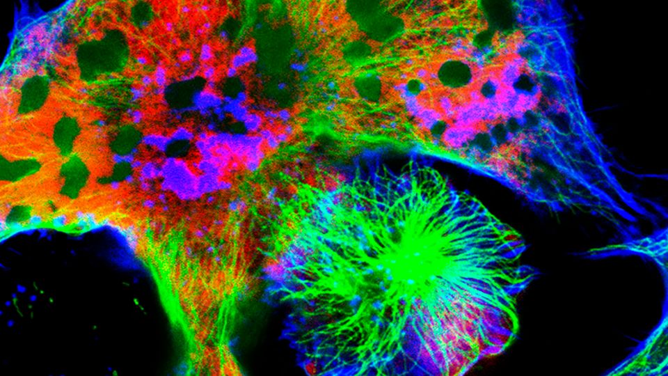
Live-cell imaging enables researchers to track subcellular dynamics in real time, revealing previously unknown cellular functions in both healthy and disease states.
Recent advancements, including AI-powered data analysis and enhanced spatiotemporal resolution, have made it an indispensable tool across basic science, diagnostics and high-throughput drug screening. However, navigating the complexities of labeling methods, imaging platforms and data processing can be daunting.
This guide provides essential strategies to streamline your live-cell imaging workflows and achieve reliable, reproducible results.
Download this guide to explore:
- Strategies for maintaining optimal cell health during long-term imaging
- Best practices for selecting labeling techniques that minimize phototoxicity
- Key considerations when choosing the right imaging platform for your research
1
How To Guide
Live-cell imaging is utilized to track subcellular dynamics, offering the chance to examine changes in cellular
structure and functions in real time.
Recent advancements in live-cell imaging technologies, including improved spatiotemporal resolution1,2 and
AI-powered data analysis for complex data sets,3 have made live-cell imaging an indispensable tool for researchers.
From applications in basic science4 to disease diagnostics5 and high-throughput drug screening,6
live-cell imaging is uncovering previously unknown cellular functions in healthy and disease states.
However, live-cell imaging technologies and methods can also be challenging to navigate, from choosing
the best labeling method to selecting an imaging platform and image processing technique. This guide will
provide tips and tricks to help guide these decisions and improve the quality and reproducibility of your livecell
imaging workflow.
Planning your live-cell research experiment
Maintaining conditions that ensure cells remain healthy and functional throughout your imaging experiment
is crucial. Imaging during periods of cellular stress, caused by environmental changes such as
fluctuations in CO2 levels or temperature, can alter experimental results. It is also important to consider
the logistics of obtaining, culturing and preparing cells for live-cell imaging. For instance, if you need to
transport live cells to another facility or location, consider using a portable cell transport carrier with
temperature control.
Careful planning is crucial for determining the timing of your imaging experiment, particularly if you are
imaging at specific time points. Diligently plan the timing of your live imaging protocol. If feasible, schedule
the imaging sessions between feeds to avoid disrupting the cells during imaging.
Labeling cells
You can label cells to target cell structures, functions and cellular proteins of interest. There are several
different options to consider:
• Fluorescent protein (FP) tagging, like green fluorescent protein (GFP), mCherry and yellow fluorescent
protein (YFP), offers high specificity because the fluorescent protein is genetically linked to the protein
of interest. This also means it is typically stable, making it suitable for long-term imaging. However,
you need to genetically edit cells to express FPs, which can be time-consuming.
Live-Cell Imaging Tips for
Experimental Success
Kaja Ritzau-Reid, PhD
LIVE-CELL IMAGING TIPS FOR EXPERIMENTAL SUCCESS 2
How To Guide
• Genetically encoded indicators are biosensors engineered from FPs that report on specific cellular
changes in real time, such as GCaMP, which reports on calcium transients, or reactive oxygen species
sensors. CRISPR/Cas9 is a popular method for genetically integrating these biosensors, enabling
minimal disruption to the cells and inducible control via specific promoters. This system has high
specificity with long-term expression.
• Chemical dyes, like silicone rhodamine (SiR) and Calcein-AM are good alternatives when genetic
tagging isn’t feasible. The dyes are typically added directly to the cell culture media, requiring membrane-
permeable delivery. Due to their ease of use, they are particularly useful for short-term imaging
and usually have good photostability.
Optimizing your labeling strategy
• Avoid over-labeling cells with fluorescent dyes as this can result in non-specific staining, increased
background signals, spectral overlap and possible cytotoxicity.
• You may want to select fluorescent dyes that are closer to the red end of the spectrum, as longer
wavelengths result in less phototoxicity.
• Avoid spectral overlaps when selecting fluorescent dyes .
• For three-dimensional (3D) live-cell cultures, such as organoids or spheroids, a minimally invasive
clearing method can improve fluorescence imaging quality.7
• Optimize your labeling method for the specific cells you are using, examining the signal-to-noise ratio
and any possible cytotoxicity effects.
Choosing the right imaging system: A trade-off between speed,
resolution and phototoxicity
Selecting the right imaging system should be guided by your experimental goals and sample characteristics.
Different imaging modalities offer various advantages and disadvantages, so knowing your priorities
is essential. Choosing an imaging platform based solely on the highest resolution may be tempting, but in
practice, this could be extremely time-consuming and unnecessary depending on your sample and goals.
Consider the following trade-offs when selecting your imaging system:
Speed vs resolution
• Fast imaging techniques such as spinning disk confocal or light sheet fluorescence microscopy
(LSFM) may sacrifice resolution to maintain cell viability. These techniques are particularly suited to
3D tissues such as organoids that require volumetric imaging.8
• Super-resolution imaging e.g., stimulated emission depletion (STED), structured illumination microscopy
(SIM), photoactivated localization microscopy (PALM) and stochastic optical reconstruction
microscopy (STORM), is typically very slow. If you aim to image nanoscale cellular structures, you will
likely prioritize resolution over speed, and these techniques may be more suitable.
Phototoxicity vs resolution
• Methods such as light sheet or confocal spinning disk have low phototoxicity and are suitable for
long-term live imaging experiments.
LIVE-CELL IMAGING TIPS FOR EXPERIMENTAL SUCCESS 3
How To Guide
• Super-resolution methods typically have high phototoxicity and can damage cells or tissue samples.
However, SIM has become popular for live imaging due to its lower phototoxicity and faster imaging
speeds compared to other super-resolution techniques.
Automated live imaging platforms for drug screening applications have become essential tools, particularly
in drug discovery and development. This is typically called high-content screening, which combines
high-resolution microscopy with automated analysis.6 This is often a costly setup with advanced
equipment. There are also more budget-friendly options for live-cell imaging in the lab. For example, it is
possible to purchase imaging devices that you can place inside the incubator to capture fluorescence and
bright field images in real time.
Keeping cells healthy
Environmental control
During live-cell imaging, it is essential to maintain the same environmental conditions for your cells that
they usually have in culture. Most cells can’t tolerate long-term temperature, pH or humidity changes.
Some cells are more sensitive than others; for example, stem cells are highly sensitive to environmental
changes, whereas some cancer cell lines may be more tolerant. Here are some tips to help ensure that
your cells stay healthy during live-cell imaging:
• For long-term live-cell imaging, ensure that your imaging platform has environmental controls set up
according to your cell culture requirements. These should include temperature, but ideally, also CO2.
• If you cannot control the CO2 levels, using a large volume of culture media will help reduce evaporation
and changes in pH.
• Consider using 4-(2-hydroxyethyl)-1-piperazineethanesulfonic acid (HEPES) buffered saline (HBS) in
your media to help maintain pH levels.
• Maintain strict aseptic technique during handling to prevent contamination. You can also consider
using antibiotics in your culture media to help prevent bacterial contamination.
• Some advanced platforms can now include integrated systems with sensing capabilities for automated
cell culture maintenance.
Minimizing phototoxicity and photobleaching
• Use the lowest possible light intensity while still producing a strong signal.
• Where possible, use fluorophores that are excited by longer wavelengths.
• Illuminate your sample for the shortest possible time.
• Reduce the frame rate as much as possible.
• Using a gentler imaging modality, such as LSFM, will help to reduce photobleaching.
• Utilizing camera features such as binning can enhance the signal-to-noise ratio. This is especially
effective in charge-coupled device (CCD) and complementary metal-oxide-semiconductor (CMOS)
cameras.
LIVE-CELL IMAGING TIPS FOR EXPERIMENTAL SUCCESS 4
How To Guide
• It is generally advisable to prioritize cell health over image resolution, which may mean using shorter
exposure times or lower magnification.
Troubleshooting tips for common problems
Cells are dying during imaging
• If you add chemical dyes for labeling, they may be causing cytotoxicity. Optimize the amount you
are using.
• Consider adding HBS to your media or using a specialized live-cell imaging solution.
• Reduce the laser intensity and exposure time.
Lots of background noise during imaging
• Check the microscope setup for misaligned light paths or dirty lenses.
• Use glass-bottom dishes, as plastic dishes can autofluoresce.
• Some media formulations will contain components that will fluoresce, causing an unwanted background
signal. You can try several options: reduce serum concentration, switch to phenol red-free
media or consider using saline solution or specialized live-cell imaging solution.
Focus drift during imaging
• Ensure the plate is stable and the system is fully equilibrated before starting the experiment.
• Use an autofocus system on your imaging platform.
Image processing and analysis
Live-cell imaging can generate massive datasets, creating a bottleneck at the data analysis stage. While
manual data analysis and correction are often still required, deep learning tools can improve efficiency
and accuracy, extracting meaningful information such as morphology and movement. Multiple opensource
tools are available to automate otherwise time-consuming processes, such as segmentation (e.g.,
CellPose 2.0 and StarDist) and cell tracking (e.g., TrackMate, DeepCell Tracking, and DeepTrack 2.0).
Future trends
Recent innovations in live-cell microscopy technology have revolutionized the field and its applications in
biomedicine. Further development in the field is on an upward trajectory, with more advanced technologies
becoming increasingly accessible. For example, multiplexing is an emerging technique in live-cell
imaging, enabling the detection of many more biomarkers than traditional fluorescence microscopy.9,10
Integrating AI and automated systems is becoming increasingly prevalent, enhancing the ability to process
and analyze large data sets and extract meaningful insights. Alongside progress in 3D in vitro tissue
culture and modeling organ-like physiology in both healthy and disease states, this is fueling the next
generation of advancements in live-cell imaging technology.
LIVE-CELL IMAGING TIPS FOR EXPERIMENTAL SUCCESS 5
How To Guide
Finally…
This is an exciting time to explore the world of live-cell imaging. Smarter technologies and AI-driven analysis
have made this a powerful and accessible technique. Take your time to plan your experiment. Carefully
consider your experimental goals when selecting your imaging platform and allow time to optimize
your cell labeling method. You probably won’t get it perfectly right the first time, but perseverance will
pay off!
References
1. Miyashiro D, Tojima T, Nakano A. Extremely high spatiotemporal resolution microscopy for live cell imaging by single
photon counting, noise elimination, and a novel restoration algorithm based on probability calculation. Front Cell Dev Biol.
2024;12:1324906. doi:10.3389/FCELL.2024.1324906/BIBTEX
2. Chin LK, Lee CH, Chen BC. Imaging live cells at high spatiotemporal resolution for lab-on-a-chip applications. Lab Chip.
2016;16(11):2014-2024. doi:10.1039/C5LC01556A
3. Shroff H, Testa I, Jug F, Manley S. Live-cell imaging powered by computation. Nat Rev Mol Cell Biol 2024 256.
2024;25(6):443-463. doi:10.1038/s41580-024-00702-6
4. Betjes MA, Zheng X, Kok RNU, van Zon JS, Tans SJ. Cell tracking for organoids: Lessons from developmental biology.
Front Cell Dev Biol. 2021;9:675013. doi:10.3389/FCELL.2021.675013/BIBTEX
5. Alieva M, Wezenaar AKL, Wehrens EJ, Rios AC. Bridging live-cell imaging and next-generation cancer treatment. Nat Rev
Cancer 2023 2311. 2023;23(11):731-745. doi:10.1038/s41568-023-00610-5
6. Esner M, Meyenhofer F, Bickle M. Live-cell high content screening in drug development. Methods Mol Biol. 2018;1683:149-
164. doi:10.1007/978-1-4939-7357-6_10,
7. Inagaki S, Nakagawa-Tamagawa N, Huynh N, et al. Isotonic and minimally invasive optical clearing media for live cell
imaging ex vivo and in vivo. bioRxiv. September 2024:2024.09.13.612584. doi:10.1101/2024.09.13.612584
8. Chen Y, Chauhan S, Gong C, et al. Low-cost and scalable projected light-sheet microscopy for the high-resolution imaging
of cleared tissue and living samples. Nat Biomed Eng 2024 89. 2024;8(9):1109-1123. doi:10.1038/s41551-024-01249-9
9. Andreou C, Weissleder R, Kircher MF. Multiplexed imaging in oncology. Nat Biomed Eng 2022 65. 2022;6(5):527-540.
doi:10.1038/s41551-022-00891-5
10. Zhanghao K, Li M, Chen X, et al. Fast segmentation and multiplexing imaging of organelles in live cells. Nat Commun 2025
161. 2025;16(1):1-14. doi:10.1038/s41467-025-57877-5
About the Author
Kaja Ritzau-Reid, PhD
Freelance Science Writer
Kaja attained a BSc in genetics, MRes in neurotechnology and a PhD in bioengineering at Imperial College
London. During her PhD, she worked on bioengineering organoids and spent part of her time working in an
organoid group in Vienna, Austria. She is currently working as a freelance science writer.